Monday, January 11, 2016

Doctors, Please Destress Yourself

By Dr Deepu

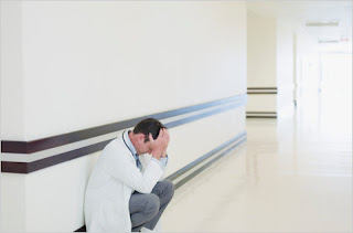
Off late there are many instances where questions on stress levels of doctors are raised. Recently a survey also revealed that a majority of resident doctors are under stress and also a startling revelation about increased suicide rates among young doctors. Here I want to share a message received on social media.

Dr.Amol Pampatwar 2011 batch Gmch Aurangabad ms Obgy died due to massive ami today at 9.20 am while travelling from miraj to Kolhapur .
He was very soft spoken person n.married a year ago .
Pm showed lad infarct with multiple plaques in coronaries.neglected chest pain for two days....
May his soul rest in peace..... Recently we are hearing news about many young doctors facing morbidity/mortality due to life style diseases like MI etc.
Here are views, though from very short experience:

Doctors are the one who neglect health and healthy lifestyle the most.
Few things that I found helpful are:
1. Practice is a continuous process... You are there in hospital for patients and not vice a versa. So dont get bothered much by fluctuations in practice.

2. Patients need you more than, you need them..
So set your limits. Stop OPD on time. Avoid patients who want to take ur leasure time (few patients feel pride in visiting u on sundays or off ur opd time).

3. 'Put your eggs in different baskets' is a good saying, but dont put them in too many baskets that you are worried about missing few. So dont keep so many attachments to hospitals.  Every new attachment demands time plus travel. Stressing us further.

4. Most important, Set your own targets ..  not the targets that ur colleague/competator has set. He may be  earning much more .. but he might be paying the cost in some other ways (family.. hobbies.. friends etc).

5. When in doubt about health  dont be your own doctor.

6. Give time to family and friends, in hour of trouble they are the one who will be with you in hospital and not ur bosses or professional colleagues. ..

7. "Happiness lies in satisfaction". If we are not satisfied, we will not be happy even if we earn highest in world.
8. Best time to be happy is now, best moment to be happy is present moment. Learn to enjoy the whole  journey, dont wait to be happy till u reach the destination!!

Monday, December 14, 2015

Nearly One-Third Of Interns, Residents May Experience Depressive Symptoms During Their Training, Analysis Suggests

The Los Angeles Times (12/9, Kaplan) reports in “Science Now” that “nearly one-third of interns and residents experience depressive symptoms or full-blown depression at some point during their training,” an analysis published Dec. 8 in the Journal of the American Medical Association suggests. After analyzing “data from 54 earlier studies involving 17,560 physicians in training,” then pooling results, investigators found that “the prevalence of depression or depressive symptoms was 29%.”
        The Washington Post (12/9, Sun) points out in “To Your Health” that “researchers said the prevalence of depression was much higher among medical residents than the general population.” In comparison, “the National Institute of Mental Health has said that in 2013, about 6.7 percent of all US adults had at least one major depressive episode during the previous year.”
        TIME (12/9, Oaklander) reports that the reasons for higher rates of depression among new physicians “are complex, ranging from long hours and sleep deprivation to bullying by attending physicians to a stigmatized attitude against mental illness.” Not only are the physicians suffering, but their patients too, as evidenced by the fact that “medical literature links untreated mental health problems in this population of young doctors to more medical errors and worse patient care.”

        According to MedPage Today (12/9, Bender), an accompanying editorial observed that even though “the medical profession is obligated to provide the best medical and mental healthcare to its members, ‘best efforts fall short, mostly because of the high levels of stigma attached to seeking mental health care.’” The editorial suggested that “perhaps an overhaul of the graduate medical education training system is in order.” 

The summary of the study is given below
The study was done to provide a summary estimate of depression or depressive symptom prevalence among resident physicians.
Information on study characteristics and depression or depressive symptom prevalence was extracted independently by 2 trained investigators. The estimates were pooled using random-effects meta-analysis. Differences by study-level characteristics were estimated using meta-regression.
The data were extracted from 31 cross-sectional studies (9447 individuals) and 23 longitudinal studies (8113 individuals). Three studies used clinical interviews and 51 used self-report instruments. The overall pooled prevalence of depression or depressive symptoms was 28.8% (4969/17 560 individuals, 95% CI, 25.3%-32.5%), with high between-study heterogeneity (Q = 1247, τ2 = 0.39, I2 = 95.8%, P < .001). Prevalence estimates ranged from 20.9% for the 9-item Patient Health Questionnaire with a cutoff of 10 or more (741/3577 individuals, 95% CI, 17.5%-24.7%, Q = 14.4, τ2 = 0.04, I2 = 79.2%) to 43.2% for the 2-item PRIME-MD (1349/2891 individuals, 95% CI, 37.6%-49.0%, Q = 45.6, τ2 = 0.09, I2 = 84.6%). There was an increased prevalence with increasing calendar year (slope = 0.5% increase per year, adjusted for assessment modality; 95% CI, 0.03%-0.9%, P = .04). In a secondary analysis of 7 longitudinal studies, the median absolute increase in depressive symptoms with the onset of residency training was 15.8% (range, 0.3%-26.3%; relative risk, 4.5). No statistically significant differences were observed between cross-sectional vs longitudinal studies, studies of only interns vs only upper-level residents, or studies of nonsurgical vs both nonsurgical and surgical residents.
The study revealed  the summary estimate of the prevalence of depression or depressive symptoms among resident physicians was 28.8%, ranging from 20.9% to 43.2% depending on the instrument used, and increased with calendar year. Further research is needed to identify effective strategies for preventing and treating depression among physicians in training.

MCI not to upload the minutes of any ethics committee meeting held


The Medical Council of India (MCI), has revised its working methodology and has decided not to make public the minutes of any ethics committee meetings, where the medical negligence cases and cases relating to professional misconduct of doctors are heard. Since October 2013, the MCI has not uploaded the minutes of any ethics committee meeting held, that is, after the current administration took over.

MCI said that the decision taken in each case would be respective to that particular case. The council further added that no consolidated minutes of the meetings of the ethics committee are now being maintained/prepared. This is done as the ethics committee of the council considers the matter before it in a quasi-judicial capacity and makes judgement upon the appeal/complaint. The committee has to decide upon the matter that affects the rights and duties of the contesting parties in accordance with the principles of natural justice and disclosing the proceedings prior to conclusion of a matter would serve no public interest.

The minutes of ethics committee meetings were uploaded regularly from May 2011 to May 2013 on the MCI website. The minutes of each committee meeting started with confirmation of the minutes of the previous meeting and all proceedings were absolutely transparent till the current council took over in November 2013.

However, some medico-legal experts have expressed their displeasure at this new turn of events and believe that minutes of hearings need to be public. It have also pointed out that this decision by MCI seems more like an attempt to hide the irregularities in the MCI that are being frequently reported. Many appeals seeking the minutes of meetings were met with stony silence till the applicants approached the chief information commissioner who had to force the MCI to hand over the specific minutes sought and threaten the public information officer with penalty.

Saturday, December 12, 2015

Chest Radiology - The Silhouette Sign

By Dr Deepu
Silhouette sign/loss of silhouette sign/ loss of outline sign.
I was always confused with the silhouette sign for its hidden meaning and failure to decode it by many medical students. So, I thought it would be apt to unravel it so that it could be handy for many medical students.
One of the most useful signs in chest radiology is the silhouette sign. This sign was described by Dr. Ben Felson. The silhouette sign is in nothing but  elimination of the silhouette or loss of lung/soft tissue interface caused by a mass or fluid in the normally air filled lung. For instance, if an intrathoracic opacity is in anatomic contact with, for example, the heart border, then the opacity will obscure that border. The sign is commonly applied to the heart, aorta, chest wall, and diaphragm. The location of this abnormality can help to determine the location anatomically. 

Just go through the X Ray to know the  various structures seen in the chest x ray.

Let me explain this with this image.
What do we see???
There is plastic bottle which is surrounded by air, the margins of the shadow is very  well demarcated from the surrounding air.

First scenario: There are two bottles, made of same material, placed apart from each other. The shadows appears separate from each other. Let us consider the right bottle to be the heart and the air surrounding the bottle as lung. The left bottle as a mass, since they are far from each other, the border of both  is visible clearly.

Second scenario: Here we see the bottles are touching each other at two points and there is no gap in between and if we look at the shadow, we cannot differentiate between the two shadows, they appear like a single opacity at the upper and lower ends.


For the heart, the silhouette sign can be caused by an opacity in the RML, lingula, anterior segment of the upper lobe, lower aspect of the oblique fissure, anterior mediastinum, and anterior portion of the pleural cavity.
This contrasts with an opacity in the posterior pleural cavity, posterior mediastinum, of lower lobes which cause an overlap and not an obliteration of the heart border. Therefore both the presence and absence of this sign is useful in the localization of pathology.

want to read more in chest radiology??? Have a look at the following pages
Chest Radiology
Signs in Chest Radiology

Thursday, September 24, 2015

Chest Radiology _ A Case Of Azygous Lobe

By Dr Deepu
The lungs are normally divided into five lobes by three main fissures .
 Occasionally, invaginations of the visceral pleura create accessory fissures that separate individual bronchopulmonary segments into accessory lobes .
 An azygos lobe is found in approximately 0.4% of patients . In contrast to other accessory lobes, the azygos lobe does not correspond to a distinct anatomical bronchopulmonary segment .

It forms during embryogenesis when the precursor of the azygos vein fails to migrate to its medial position in the mediastinum, where it normally arches over the origin of the right upper lobe bronchus.
 This gives rise to the following characteristics, which are visible on a standard chest x-ray

: the laterally displaced azygos vein lies between folds of parietal pleura, also referred to as the mesoazygos, where it assumes a characteristic teardrop shape ; the mesoazygos indents the right upper lobe, thereby creating the accessory (azygos) fissure, which is similar in shape to an inverted comma; the fissure delineates the azygos lobe, located superomedially ; laterally, the pleural folds of the mesoazygos separate before reaching the chest wall, resulting in a radiopaque triangular area ; and medially, the tracheobronchial angle appears empty .


 An azygos lobe may be confused with a pathological air space such as a bulla or abscess . In addition, the abnormally located azygos vein may be mistaken for a pulmonary nodule, while a consolidated azygos lobe may be confused with a mass . An understanding of the pathogenesis and characteristic x-ray features of the azygos lobe will enable an accurate diagnosis in most cases .
 If the x-ray findings are equivocal, computed tomography will be diagnostic .

Saturday, September 12, 2015

Sleep-Disordered Breathing In Kids May Be Linked To Academic Problems


By Dr Deepu

HealthDay (9/9) reports that research suggests that “when children have sleep troubles due to breathing problems – such as sleep apnea – they may struggle in school.” Investigators “reviewed the results of 16 studies dealing with sleep apnea or related disorders in children and academic achievement.” The data indicated that kids “with sleep-disordered breathing did worse in language arts, math and science tests compared to those without such conditions.” The findings were published online in Pediatrics.
The researchers reviewed the results of 16 studies dealing with sleep apnea or related disorders in children and academic achievement. The investigators found that children with sleep-disordered breathing did worse in language arts, math and science tests compared to those without such conditions.


Chest Medicine Made Easy -By Dr Deepu : BASICS OF CHEST X RAY- PART 2 , EASY WAY TO INTERP...

Chest Medicine Made Easy -By Dr Deepu : BASICS OF CHEST X RAY- PART 2 , EASY WAY TO INTERP...: Welcome to basics of CXR, this is part two of this series, read my previous post part 1, in the next post we will be exploring about the s...

By Dr Deepu
Related Posts Plugin for WordPress, Blogger...